
Fundus Examination: The Window into Your Eye Health That Everyone Should Understand
Somewhere behind your pupil lies a hidden landscape — a terrain of blood vessels, nerve fibres, and light-sensitive tissue that reveals secrets about your health no other part of your body can show. This is the fundus, and examining it provides insights not just into your eye health, but into conditions affecting your entire body.
A fundus examination is one of the most valuable yet underappreciated tests in medicine. It's the only place in the body where blood vessels and nerve tissue can be observed directly, without surgery or invasive procedures. Understanding what this examination involves and why it matters empowers you to take control of your health. The best eye specialist in Indirapuram performs thorough fundus examinations that can detect problems before symptoms ever appear.
What Is a Fundus Examination?
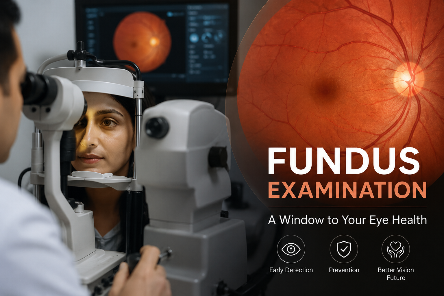
The fundus refers to the interior back surface of the eye — the retina, macula, optic disc (where the optic nerve enters the eye), and blood vessels that supply these structures. A fundus examination uses specialised instruments to view and assess these tissues.
The examination typically involves pupil dilation using eye drops. Dilation widens the pupil, allowing your eye specialist to see a much larger area of the fundus. While undilated examination is possible with some modern instruments, dilation provides the most comprehensive view.
Several techniques may be used: direct ophthalmoscopy (a handheld instrument providing a magnified view), indirect ophthalmoscopy (providing a wider field of view), slit-lamp biomicroscopy with special lenses, and fundus photography (creating detailed images for documentation and comparison).
What the Fundus Reveals
Diabetic Retinopathy: Diabetes damages small blood vessels throughout the body, and the retinal vessels are no exception. Fundus examination reveals characteristic changes — microaneurysms, haemorrhages, exudates, and in advanced cases, abnormal new vessel growth. Detecting these changes early allows intervention before vision loss occurs.
Hypertensive Retinopathy: High blood pressure affects retinal arteries, causing them to narrow, leak, or show characteristic crossing changes where arteries and veins intersect. The fundus often reveals hypertension before patients experience symptoms, and it shows whether blood pressure control is adequate.
Glaucoma: The optic disc appearance changes characteristically in glaucoma, with increased cupping and nerve fibre layer loss. Fundus examination allows monitoring of these changes over time, tracking whether the disease is stable or progressing despite treatment.
Macular Degeneration: Age-related macular degeneration shows characteristic drusen (yellow deposits), pigment changes, and in wet AMD, abnormal blood vessel growth and fluid accumulation. Early detection allows treatment that can preserve central vision.
Retinal Detachment Risk: Peripheral retinal tears, lattice degeneration, and other predisposing conditions are visible during dilated fundus examination. Identifying these risks allows preventive treatment before detachment occurs. The best eye doctor in Indirapuram performs a thorough peripheral retinal assessment.
Beyond Eye Disease: Systemic Conditions
The fundus examination's value extends beyond ophthalmology. Because retinal blood vessels are visible directly, they provide a window into vascular health throughout the body.
Cardiovascular risk: Retinal vessel changes correlate with cardiovascular disease risk. Narrowed arteries, arteriovenous nicking, and other changes suggest systemic vascular damage that may affect the heart and brain.
Brain tumours: Increased intracranial pressure causes papilledema — swelling of the optic disc visible during fundus examination. This finding may be the first indication of a brain tumour or other space-occupying lesion.
Multiple sclerosis: Optic neuritis, visible as optic disc changes, is often an early sign of MS. Fundus examination combined with patient symptoms helps guide neurological evaluation.
Blood disorders: Conditions like leukaemia, anaemia, and sickle cell disease produce characteristic retinal findings. The fundus can reveal these conditions or suggest the need for blood testing.
Who Needs Regular Fundus Examination?
Everyone over 40: Age-related eye conditions become increasingly common. Annual dilated examinations catch problems early.
Diabetics: Regardless of age, diabetics need an annual fundus examination. If retinopathy is detected, more frequent monitoring is essential.
Hypertensives: Regular fundus examination monitors end-organ damage and treatment effectiveness.
High myopes: Severe nearsightedness increases the risk of retinal detachment. Peripheral retinal examination is particularly important.
Family history of eye disease: Glaucoma, macular degeneration, and some retinal conditions have genetic components. Earlier and more frequent screening is advisable.
Due for your fundus examination? Contact the eye care clinic near Indirapuram at 98999 60700.
What to Expect During the Examination
The examination itself is painless. Dilating drops take 20-30 minutes to work fully. During this time, you may experience mild stinging and gradually increasing light sensitivity. Once dilated, your eye specialist examines the fundus using various instruments, each providing different views and magnifications.
After the examination, pupils remain dilated for 4-6 hours. During this time, near vision is blurry and light sensitivity is increased. Bring sunglasses and arrange transportation — driving immediately after dilation isn't advisable. By evening, vision typically returns to normal.
Fundus photography may be performed, creating permanent images for comparison over time. These photos become part of your medical record, documenting the baseline against which future changes are measured.
Frequently Asked Questions
Is dilation really necessary?
For a comprehensive examination, yes. An undilated pupil is like looking through a keyhole — you see a small central area but miss the periphery. Many important findings, particularly retinal tears and diabetic changes, occur in peripheral areas only visible with dilation.
How often should I have a fundus examination?
Annual examination is standard for adults over 40 and anyone with diabetes, hypertension, or a family history of eye disease. Younger adults without risk factors may go longer between examinations. The best eye specialist in Indirapuram recommends intervals based on your individual risk factors.
Can I drive after dilation?
It's not recommended. Dilation causes light sensitivity and blurred near vision that can impair driving safety. Arrange alternative transportation or plan to wait several hours before driving.
What if something abnormal is found?
Many findings require only monitoring. Others need treatment or further investigation. Your specialist will explain any findings, their significance, and recommended next steps. Early detection generally means more treatment options and better outcomes.
Where can I get a comprehensive fundus examination?
Samyak Eye Care offers thorough fundus examinations with advanced imaging technology. The eye care clinic near Indirapuram serves patients from Vaishali, Kaushambi, Surya Nagar, and throughout Ghaziabad. Visit our clinic for your examination.
Does fundus examination hurt?
No. The dilating drops may cause mild, brief stinging. The examination itself involves bright lights that can be uncomfortable but cause no pain. Most patients find the temporary light sensitivity afterwards more bothersome than the examination itself.
See Beyond the Surface
A fundus examination takes just minutes but reveals information that could protect your vision — and your life — for decades. It's a simple, painless test with profound implications for early detection and prevention. Whether you're managing diabetes, monitoring glaucoma, or simply maintaining routine eye health, this examination provides irreplaceable insights into what's happening inside your eyes.
SCHEDULE YOUR FUNDUS EXAMINATION TODAY
Call: 98999 60700
Website: www.samyakeyecare.com
Location: Gaur Heights, Sector 4, Vaishali, Ghaziabad
Serving patients from Indirapuram | Vaishali | Kaushambi | Surya Nagar | Ghaziabad


