
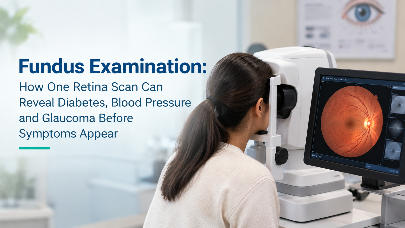
Fundus Examination: How One Retina Scan Can Reveal Diabetes, Blood Pressure and Glaucoma Before Symptoms Appear
There is a moment in a fundus examination that never loses its clinical weight. The pupil dilates, the light enters, and the retina comes into view. Blood vessels, optic disc, macula, peripheral retina, all visible through a non-invasive instrument without a single incision or blood draw. In that 10-minute examination, conditions that may have been silently progressing for years become visible. Not theoretically visible. Actually, directly, immediately visible.
A 52-year-old businessman from Ghaziabad came to Samyak Eye Care Clinic for what he described as a routine glasses check. He had known about his type 2 diabetes for four years and attended regular check-ups with his physician. His HbA1c was reasonably controlled. He felt well. His previous eye examinations at a general clinic had checked his prescription and nothing else. His fundus examination at our clinic revealed moderate non-proliferative diabetic retinopathy in both eyes, with microaneurysms, dot haemorrhages, and early hard exudates. Changes that had been accumulating silently while his prescription was being updated and his general health appeared managed.
He was referred promptly for laser treatment. His vision is intact. But the examination that caught his retinopathy was not the examination he had been attending. It was the first time anyone had looked at the back of his eyes properly.
As the best eye specialist in Vaishali, Ghaziabad, I want to explain clearly what a fundus examination reveals, why it reveals it earlier than most other investigations, and which patients in Ghaziabad and across NCR need one immediately.
What a Fundus Examination Actually Shows
The retina is the only place in the human body where blood vessels can be directly visualised without any surgical procedure. When those blood vessels are examined, they reveal not just the state of the retina but the state of the circulatory system more broadly. The microvascular changes visible in the retina mirror the microvascular changes occurring in the kidneys, the heart, and the brain in response to systemic diseases like diabetes and hypertension.
A fundus examination provides direct visual access to:
The retinal blood vessels: Their calibre, regularity, branching pattern, and whether they show signs of leakage, haemorrhage, or abnormal new vessel growth
The optic disc: The entry point of the optic nerve into the eye, where the characteristic cupping and pallor of glaucoma is visible before visual field loss becomes symptomatic
The macula: The central retinal region responsible for fine detail vision, where swelling, deposits, and degeneration affect central vision
The peripheral retina: Where retinal tears, early detachment, and peripheral vascular changes can be identified before they cause symptoms
What Fundus Examination Reveals About Systemic Health
Diabetic retinopathy
Research published in ophthalmology literature found that among individuals with a known history of diabetes who underwent fundus examination, 54.83 per cent showed detectable fundus lesions. More than half. In India, where the International Diabetes Federation estimates over 100 million adults are living with diabetes, a significant proportion of whom have inadequate access to structured retinal screening, this figure represents an enormous burden of undetected retinal disease.
Diabetic retinopathy progresses through defined stages. In the early non-proliferative stages, there are no symptoms. The patient sees normally. The retinopathy is visible on fundus examination and treatable with good outcome. In the advanced proliferative stage, abnormal new blood vessels have grown across the retina and vitreous, and the risk of sudden, severe, and potentially irreversible vision loss is high. The difference between these stages is years. The opportunity to intervene is entirely in the earlier stages. The fundus examination is the tool that identifies which stage a patient is at.
Hypertensive retinopathy
Among individuals with a known history of hypertension who underwent fundus examination in a large screening study, 54.06 per cent showed positive fundus findings. The retinal blood vessel changes of hypertension, including arteriovenous nicking, copper or silver wiring of the arteries, and in severe cases, flame-shaped haemorrhages and papilloedema, are direct windows into the systemic vascular state.
Research confirms that hypertensive retinopathy not only signals existing eye problems but may indicate serious systemic vascular disease, including the elevated risk of stroke or heart attack. A fundus examination that reveals significant hypertensive changes in a patient who was not aware their blood pressure was poorly controlled can trigger a management intervention that protects not just their vision but their cardiovascular health.
Glaucoma
Glaucoma is often called the silent thief of sight because it destroys peripheral vision gradually and painlessly, with no symptoms until a significant portion of vision is already lost. The optic disc changes of glaucoma, characterised by progressive cupping and rim thinning, are visible on fundus examination before visual field loss is detectable on standard perimetry in many cases.
A North India study published in the Indian Journal of Ophthalmology found that 53.9 per cent of glaucoma patients had one or more systemic risk factors, with hypertension present in 38.9 per cent and diabetes in 23.9 per cent. Many of these patients would benefit from fundus examination both for glaucoma assessment and for systemic disease monitoring.
Retinal detachment and peripheral tears
A retinal tear can exist silently for weeks or months before causing a symptomatic detachment. The symptoms of retinal detachment (flashing lights, a shower of floaters, a dark curtain in the visual field) are emergency signals that the tear has already progressed. During a dilated fundus examination, peripheral retinal tears and areas of lattice degeneration can be identified and treated prophylactically with laser before detachment occurs. This is one of the clearest examples of an examination finding a problem before the patient has any symptoms of it.
Who Needs a Fundus Examination and How Often
At Samyak Eye Care Clinic, I recommend fundus examination for the following groups, at minimum:
Every person with diabetes: Annual fundus examination from the time of diabetes diagnosis, regardless of how well the diabetes appears controlled. Retinopathy can develop even in patients with good HbA1c control.
Every person with hypertension: At diagnosis and annually, as hypertensive retinal changes provide direct information about systemic vascular control.
Every person over 40: Annual fundus examination as part of comprehensive eye care, to screen for early glaucoma, age-related macular changes, and retinal vascular pathology.
Every person with high myopia (above -5D): Regular fundus examination because high myopia significantly elevates lifetime risk of retinal tears, detachment, and myopic maculopathy.
Anyone with a family history of glaucoma or retinal detachment: Annual examination from age 35 or earlier depending on risk factors.
Anyone who has experienced floaters, flashes, or any visual disturbance: Urgent fundus examination on the same day or within 24 hours.
What the Examination Involves
A fundus examination at Samyak Eye Care Clinic begins with dilation of the pupil using eye drops. The dilation process takes 20 to 30 minutes to reach full effect. During this time, the patient will notice increasing sensitivity to light and blurring of near vision. These effects are temporary, lasting 4 to 6 hours for most patients.
Once the pupil is dilated, the examination itself is rapid. Using a slit lamp with a specialised fundus lens, or an indirect ophthalmoscope for peripheral retinal assessment, I examine all zones of the fundus systematically. The examination is entirely non-invasive and painless. Where indicated, fundus photography is performed to document the findings and enable comparison at future visits, which is particularly important for monitoring diabetic retinopathy progression or glaucomatous optic disc changes.
Patients should arrange not to drive immediately after the examination, as the dilating drops affect near vision and light sensitivity. Bringing sunglasses for the journey home is advisable.
Frequently Asked Questions
Q: What is a fundus examination?
A: A fundus examination is a dilated eye examination that allows direct visualisation of the retina, optic nerve, and retinal blood vessels. It detects conditions including diabetic retinopathy, hypertensive retinopathy, glaucoma, macular degeneration, retinal tears, and detachment, many of which are entirely without symptoms in their early, treatable stages.
Q: Why do the eyes need to be dilated for a retina check?
A: Pupil dilation with eye drops temporarily enlarges the pupil, allowing a much wider view of the retina, particularly the peripheral zones where retinal tears and early detachment can occur. Without dilation, only the central retina is visible. Full peripheral retinal examination requires a dilated pupil.
Q: How often should a diabetic get a fundus examination?
A: Every diabetic patient should have a fundus examination annually from the time of diabetes diagnosis. If diabetic retinopathy is detected, the frequency increases based on the severity of the findings. Research shows that more than 54 per cent of diabetics show fundus changes on examination, making annual review a clinical necessity, not an optional extra.
Q: Is a fundus examination painful?
A: No. The examination is entirely non-invasive. The dilating drops cause temporary light sensitivity and blurring of near vision, but no pain. The examination itself takes 10 to 15 minutes. Patients should expect to spend 45 to 60 minutes at the clinic total, including the waiting period for the drops to take effect.
Q: Where can I get a fundus examination in Ghaziabad?
A: Samyak Eye Care Clinic in Vaishali, Ghaziabad provides comprehensive fundus examination including fundus photography and systematic retinal assessment. We serve patients from across Ghaziabad, Vaishali, Indirapuram, Kaushambi, Surya Nagar, and NCR. Visit our clinic to book your appointment.
The Examination That Changed What Was Possible
The businessman from Ghaziabad attends his annual fundus examination now without prompting. His retinopathy has been stable for eighteen months following laser treatment. He told me at his last visit that he had mentioned the examination to his brother, who also has diabetes, and his brother had never had a retinal check. He sent his brother to us. His brother also had early retinopathy that had not been caught.
Two examinations. Two early diagnoses. Two patients whose vision is protected not because of dramatic intervention but because someone looked at the back of their eyes properly. If you have diabetes, hypertension, high myopia, or are over 40 and have not had a dilated fundus examination in the past year, visit our clinic at Samyak Eye Care, Vaishali, Ghaziabad.
Book Your Fundus Examination in Ghaziabad Today
Dr. Shalini Jain | Samyak Eye Care Clinic | Vaishali, Ghaziabad
Call: 98999 60700


