
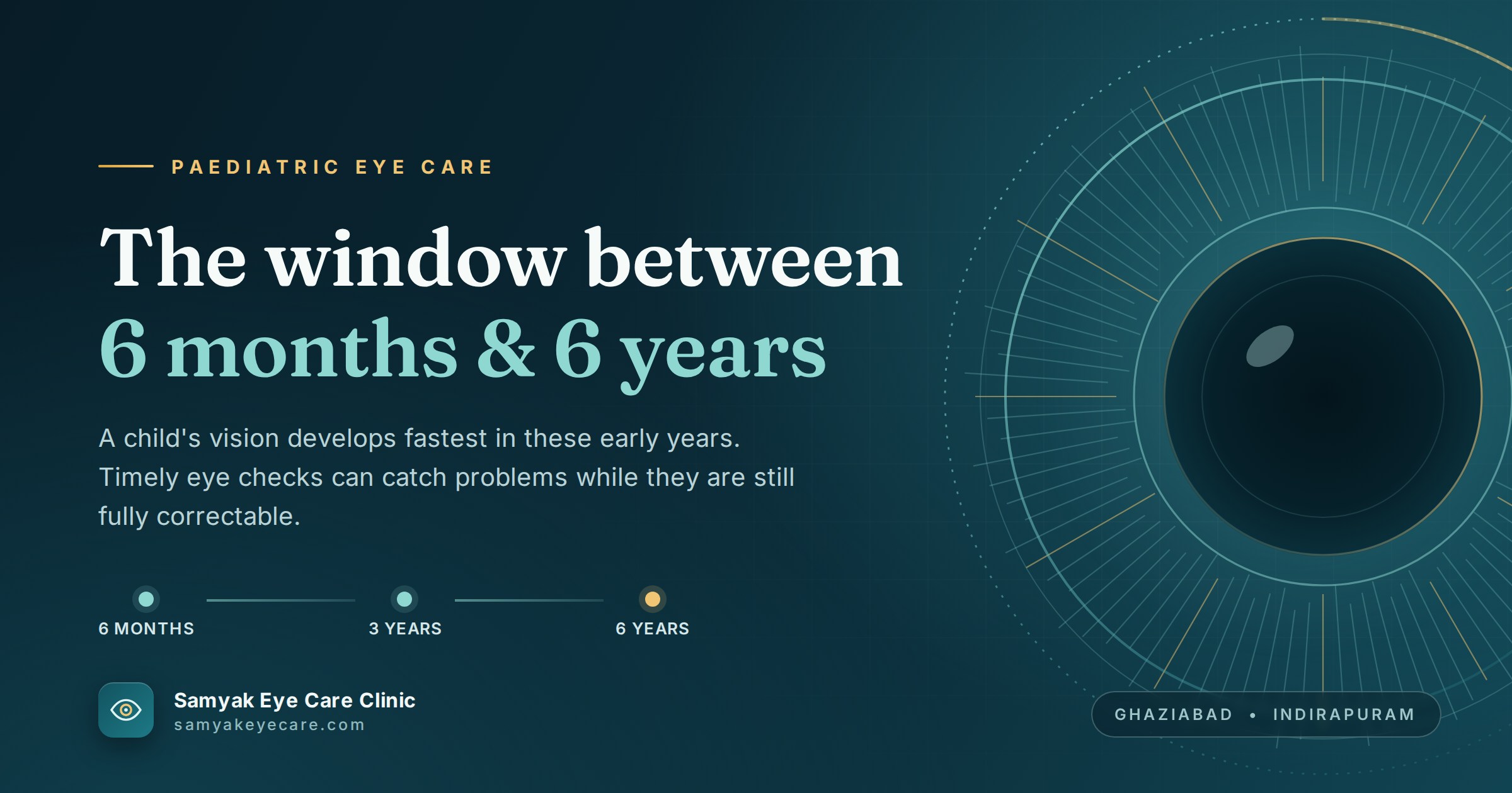
Eye Care for Children from 6 Months to 6 Years: The Window Most Ghaziabad Parents Do Not Know Exists
Most parents in Ghaziabad bring their children for a first eye examination when a teacher flags a concern, or when the child is old enough to articulate that they cannot see the board clearly. Both of these triggers place the first examination at school age, typically between 5 and 8 years old. For the conditions I am about to describe, school age is not early. In several cases, it is too late for the best possible outcome.
This is not a theoretical concern. The Indian Journal of Ophthalmology's guidelines on paediatric eye screening, published in a peer-reviewed consensus statement, identify the period from birth to 6 years as the most clinically consequential window in a child's visual life. The conditions that cause the most permanent vision loss in Indian children are not the ones that develop in adolescence. They are the ones that are present from birth or early infancy and that have strict biological timelines for intervention that most parents are never told about.
As the best eye doctor in Vaishali, Ghaziabad, with over 25 years of paediatric eye care experience, I want every parent in Ghaziabad, Indirapuram, Kaushambi, and Surya Nagar to know what the early window contains, what it requires, and why it cannot be recovered once it closes.
The Conditions That Are Present Before a Child Can Tell You Something Is Wrong
Congenital Cataract: A Six-Week Countdown
Congenital cataract, a clouding of the lens present at or shortly after birth, affects approximately 1 in 2,000 newborns globally. In India, where access to structured newborn eye screening remains inconsistent outside major hospitals, the diagnosis is frequently delayed.
The clinical timeline is unforgiving. Research published in the British Medical Journal documents that irreversible visual cortex changes begin at approximately 6 weeks of life for unilateral congenital cataract, and around 10 weeks for bilateral cataract. Evidence from long-term follow-up studies indicates an average loss of one Snellen visual acuity line for every three weeks of surgical delay during the first 14 weeks of life.
What this means in plain terms: a unilateral congenital cataract identified and treated at 4 weeks of age has an excellent visual prognosis. The same cataract identified at 4 months, because the family was waiting for the next general check-up, may result in permanent amblyopia in that eye that treatment will not fully reverse.
The warning sign parents can check at home: a white or cloudy appearance in the pupil (leucocoria), visible especially in flash photographs where a white reflection appears instead of the normal red-eye effect. Any parent who sees this in a photograph should seek urgent ophthalmological assessment, not a scheduled appointment at the next available date.
Retinoblastoma: The 90 Per Cent Statistic Parents Need to Hear
Retinoblastoma is a malignant tumour of the retina, affecting approximately 1 in 16,000 to 18,000 live births. It is the most common intraocular malignancy of childhood globally and accounts for a significant proportion of childhood cancer cases in India.
Here is the statistic that every parent of a young child needs to know: the cure rate for retinoblastoma when identified early is 90 per cent or better. When identified late, after the tumour has extended beyond the eye, outcomes are dramatically worse. Delayed diagnosis, due to missed leucocoria on red reflex testing or delayed specialist referral, is one of the most significant preventable causes of both vision loss and mortality in Indian children with this condition.
A case report published in a peer-reviewed ophthalmology journal documented a child whose retinoblastoma was initially misdiagnosed as congenital cataract at a primary healthcare centre, with specialist referral delayed by three months due to financial constraints. This pattern of delayed diagnosis, misdiagnosis, and delayed referral is not unusual in the Indian clinical context.
The red reflex test, performed using a direct ophthalmoscope, is the standard screening tool for retinoblastoma in infants. It should be performed at birth, at 6 to 8 weeks, and at every paediatric check-up in the first two years of life. Any absent or abnormal red reflex requires urgent ophthalmological referral, the same day where possible.
Flash photography can assist parents between appointments. A white or yellow reflection from one or both pupils in a photograph, the absence of the normal red-eye effect, is a reason for immediate assessment.
Retinopathy of Prematurity: A Condition That Moves Quickly
Retinopathy of Prematurity (ROP) affects the blood vessels of the immature retina in premature infants. Indian studies have reported ROP incidence of 20 to 22 per cent in screened preterm newborns, a significant proportion given India's large birth cohort. Given the growth in neonatal survival rates for premature infants, the number of children at risk of ROP in India is increasing.
The National Health Mission guidelines for ROP in India specify screening for all infants born before 34 weeks of gestation or weighing under 2,000 grams at birth, with the first examination at 4 weeks of postnatal age or 31 weeks post-conception, whichever is later. This is not optional, and it is not routine. It requires a specific ophthalmological examination by a trained specialist.
Parents of premature infants who are discharged from the NICU must ensure their infant receives the ROP examination on schedule. The condition can progress to retinal detachment within days to weeks in its most severe forms. Early laser treatment or anti-VEGF injection can prevent progression to blindness. Missed screening means missed treatment opportunity.
Infantile Glaucoma: Identifiable by Symptoms Parents Describe as Something Else
Infantile glaucoma (raised intraocular pressure from birth or early infancy) produces symptoms that parents frequently attribute to other causes. The classic triad: excessive tearing (often assumed to be a blocked tear duct), sensitivity to light (often attributed to photosensitivity), and an enlarged or cloudy cornea (often attributed to the eye simply looking large or beautiful).
Infantile glaucoma is rare but rapidly destructive. Untreated, it causes permanent optic nerve damage and visual field loss within weeks to months. It requires surgical management and the prognosis is closely tied to how early the diagnosis is made.
Any infant or toddler with persistent watery eyes that do not respond to blocked tear duct treatment, combined with light sensitivity and eyes that appear unusually large, should be assessed by a paediatric eye specialist in Ghaziabad promptly.
The Conditions That Develop Gradually in the Early Years
Amblyopia: Eight Years to Act
Amblyopia develops when the visual cortex fails to establish normal neural connections with one or both eyes during the critical developmental period. The most common causes are uncorrected significant refractive errors, strabismus, and visual deprivation from conditions like cataract or ptosis.
The treatment window for amblyopia (lazy eye) is not unlimited. The visual cortex retains the plasticity necessary for treatment up to approximately 7 to 8 years of age. Before this point, patching, atropine penalisation, and vision therapy can produce excellent outcomes. After this window closes, treatment produces limited improvement regardless of how consistently it is applied. The amblyopia that exists at age 9 in an untreated child will largely persist into adulthood.
The practical implication: every year between ages 1 and 7 that amblyopia goes undetected is a year of vision development that cannot be recovered. A comprehensive eye examination at 3 years of age specifically targets amblyopia detection while the treatment window is wide open.
Strabismus: Not Something Children Grow Out Of
Intermittent or constant strabismus (squint) in young children is frequently minimised by well-meaning family members who suggest the child will grow out of it. This is true only for a very narrow category of neonatal strabismus in the first weeks of life. For the vast majority of childhood strabismus, waiting leads directly to amblyopia through suppression, compounding one manageable condition into two.
Any squint noticed in a child over 3 months of age should be assessed by a paediatric eye specialist. There is no clinical argument for waiting to see if it resolves independently.
The Schedule Ghaziabad Parents Should Follow
Based on Indian Journal of Ophthalmology guidelines and clinical evidence, the recommended examination schedule for children from birth to 6 years is:
Birth (or within the first week): Red reflex testing by the paediatrician or neonatologist. If abnormal or difficult to assess, same-day specialist referral.
4 weeks (for preterm infants under 34 weeks gestation or under 2kg birth weight): ROP screening by a trained ophthalmologist. This is non-negotiable for eligible infants.
6 to 8 weeks: Red reflex repeat and external inspection of the eye by the paediatrician at the routine immunisation visit. Any abnormality: urgent ophthalmology referral.
6 months: First comprehensive paediatric eye examination. All findings detailed in the step-by-step guide above. This is the first opportunity to identify significant refractive errors, strabismus, early amblyopia risk factors, and structural anomalies.
3 years: Second comprehensive examination. Accurate amblyopia assessment is now possible. The treatment window for amblyopia and strabismus remains wide open.
5 to 6 years (before school entry): Third examination. The visual system must be ready for the sustained near work that school demands. Undetected refractive errors at this stage directly compromise academic start.
At our clinic, I adjust this schedule individually based on risk factors identified at each visit. A child with a family history of retinoblastoma or glaucoma follows a more intensive schedule from birth.
How to Check at Home Between Appointments
Between professional examinations, parents can perform two simple checks at home:
The flash photograph test
Take a flash photograph of your child in a slightly darkened room. Both pupils should show a symmetrical red or orange reflex. A white, yellow, or absent reflex in one or both eyes requires urgent professional assessment. This test has identified retinoblastoma and congenital cataract in children whose parents simply took a family photograph.
The eye covering test
Cover each eye alternately with your hand while the child is looking at something they enjoy. A child with good vision in both eyes will generally tolerate this without objection. A child who objects strongly to one eye being covered but tolerates the other may be indicating that the uncovered eye does not see well and they rely on it. This is not diagnostic, but it is a prompt to seek professional assessment.
Frequently Asked Questions
Q: When should a baby have their first eye examination?
A: Red reflex testing should occur at birth and again at 6 to 8 weeks as part of routine infant health checks. A comprehensive paediatric eye examination is recommended at 6 months. Premature infants born before 34 weeks or under 2kg require ROP screening at 4 weeks of postnatal age, regardless of other circumstances.
Q: What are the signs of eye problems in infants and toddlers?
A: White or yellow pupil in photographs, persistent eye watering unresponsive to treatment, sensitivity to light, eyes that appear unusually large, visible eye turn, excessive eye rubbing, or a child who objects to one eye being covered. Any of these requires prompt professional assessment, not a wait-and-see approach.
Q: Can a baby's eye condition be treated effectively?
A: Yes, and the earlier the better. Congenital cataract treated surgically in the first 6 to 10 weeks of life can achieve good visual outcomes. Amblyopia treated before age 7 to 8 shows excellent response. Retinoblastoma detected early has a cure rate of 90 per cent or better. The outcomes for all these conditions drop measurably with each week of delayed diagnosis.
Q: Is the 6-month eye examination necessary if my baby seems to see fine?
A: Yes. Congenital cataract, significant refractive errors, early amblyopia risk factors, and structural eye anomalies produce no obvious symptoms visible to a parent. A child with a significant prescription in one eye appears entirely normal in daily life because the other eye compensates. The 6-month examination uses objective techniques that require no cooperation from the infant and detect what observation cannot.
Q: Where can I get my infant's eyes examined in Ghaziabad?
A: Samyak Eye Care Clinic in Vaishali, Ghaziabad offers comprehensive paediatric and infant eye examinations from birth onwards. Our examinations for infants use objective techniques requiring no verbal cooperation. Visit our clinic to schedule your baby's first examination.
The Window Is Open Right Now
If your child is between 6 months and 6 years old and has not had a comprehensive eye examination, the window is open right now. Not indefinitely. The biological timelines for the conditions described in this blog do not wait for a convenient appointment slot or for the child to start school.
Samyak Eye Care Clinic in Vaishali has been providing paediatric eye care in Ghaziabad for over 25 years. We have examined children from the first weeks of life. We use child-adapted techniques at every age. And we have seen firsthand what early diagnosis does for a child's visual future. Visit our clinic and use the window while it is still open.
Book Your Child's First Eye Examination in Ghaziabad
Dr. Shalini Jain | Samyak Eye Care Clinic | Vaishali, Ghaziabad
Call: 98999 60700


